Embark on a journey into the fascinating world of dermatology with our comprehensive label skin diagram worksheet answers. This guide delves into the intricate structure and vital functions of the skin, unraveling its remarkable role in safeguarding and sustaining our bodies.
Prepare to uncover the secrets of the skin’s protective barrier, its thermoregulatory prowess, and its sensory capabilities. As we peel back each layer of the epidermis, dermis, and hypodermis, you will gain a profound understanding of the skin’s anatomy and its pivotal role in our overall well-being.
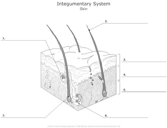
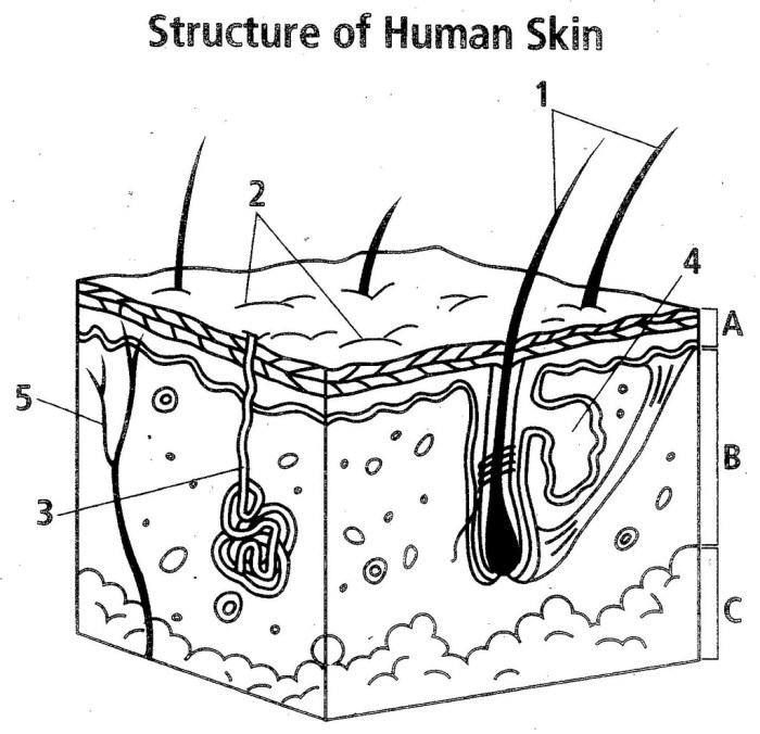
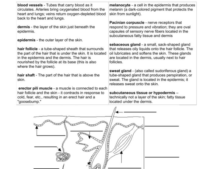
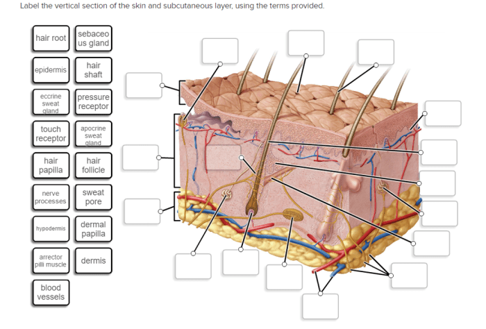
Label the Skin Diagram

The skin is the largest organ of the human body, serving as a protective barrier against external threats while maintaining homeostasis and facilitating sensory perception. It consists of three distinct layers: the epidermis, dermis, and hypodermis.
The outermost layer, the epidermis, is composed primarily of keratinized cells and serves as a waterproof barrier. Beneath the epidermis lies the dermis, which contains blood vessels, hair follicles, sweat glands, and connective tissue. The innermost layer, the hypodermis, consists of fat cells and provides insulation and cushioning.
Epidermis
- Stratum corneum: The outermost layer of dead, keratinized cells that provides a waterproof barrier.
- Stratum lucidum: A thin, translucent layer found only in thick skin, such as the palms and soles.
- Stratum granulosum: Contains cells that produce keratin, a protein that strengthens the skin.
- Stratum spinosum: Contains cells with spiny projections that connect to neighboring cells.
- Stratum basale: The deepest layer of the epidermis, containing stem cells that divide to produce new skin cells.
Dermis
- Papillary layer: The upper layer of the dermis, containing blood vessels, nerve endings, and hair follicles.
- Reticular layer: The lower layer of the dermis, containing collagen and elastin fibers that provide strength and elasticity.
- Sweat glands: Produce sweat to regulate body temperature.
- Hair follicles: Produce hair shafts that protect the body from the elements.
- Blood vessels: Supply nutrients and oxygen to the skin.
Hypodermis
- Adipocytes: Fat cells that provide insulation and cushioning.
- Connective tissue: Supports and connects the skin to underlying structures.
- Nerve endings: Transmit sensory information to the brain.
Explain the Functions of the Skin
The skin, the largest organ of the human body, serves a multitude of essential functions. It acts as a protective barrier, regulating body temperature, and facilitating sensory perception.
Protective Functions, Label skin diagram worksheet answers
The skin’s outermost layer, the epidermis, forms a waterproof barrier that shields the body from external threats. It prevents the entry of pathogens, such as bacteria and viruses, and protects against harmful UV radiation from the sun.
Thermoregulatory Functions
The skin plays a crucial role in maintaining body temperature. When the body temperature rises, the skin’s blood vessels dilate, allowing more blood to flow near the skin’s surface, where heat can be dissipated through sweating. Conversely, when the body temperature drops, the blood vessels constrict, reducing blood flow to the skin’s surface and conserving heat.
Sensory Functions
The skin contains numerous sensory receptors that allow us to perceive the world around us. These receptors enable us to detect touch, temperature, and pain. The skin also contains specialized receptors that respond to specific stimuli, such as pressure, vibration, and itching.
Describe the Structure of the Epidermis

The epidermis is the outermost layer of the skin and acts as a protective barrier against external threats. It is composed of keratinized cells, which are tough and waterproof, and arranged in multiple layers.
Layers of the Epidermis
The epidermis consists of four distinct layers:
- Stratum Corneum:The outermost layer, composed of dead, flattened cells filled with keratin, providing a waterproof barrier.
- Stratum Lucidum:Found only in thick skin, this thin layer contains cells with a high concentration of lipids, enhancing the skin’s waterproofing ability.
- Stratum Granulosum:Composed of cells containing granules of a protein called keratohyalin, which helps form the protective barrier.
- Stratum Basale:The innermost layer, containing living cells that divide and produce new cells to replace those lost from the outer layers.
Describe the Structure of the Dermis
The dermis is the middle layer of the skin, located beneath the epidermis and above the subcutaneous layer. It is composed of connective tissue, blood vessels, nerves, and hair follicles. The dermis provides support and elasticity to the skin and protects the body from external damage.The
main structural components of the dermis are collagen and elastin fibers. Collagen fibers are strong, inelastic fibers that provide tensile strength to the skin. Elastin fibers are elastic fibers that allow the skin to stretch and recoil. The dermis also contains a network of blood vessels that supply nutrients to the skin and remove waste products.
Nerves in the dermis transmit sensory information to the brain.The dermis is divided into two layers: the papillary layer and the reticular layer. The papillary layer is the upper layer of the dermis and is composed of loose connective tissue.
The reticular layer is the lower layer of the dermis and is composed of dense connective tissue.
Blood Vessels in the Dermis
The dermis contains a network of blood vessels that supply nutrients to the skin and remove waste products. The blood vessels in the dermis are arranged in two layers: the superficial vascular plexus and the deep vascular plexus. The superficial vascular plexus is located in the papillary layer of the dermis and is composed of small blood vessels that supply nutrients to the epidermis.
The deep vascular plexus is located in the reticular layer of the dermis and is composed of larger blood vessels that supply nutrients to the dermis and subcutaneous layer.
Nerves in the Dermis
The dermis contains a network of nerves that transmit sensory information to the brain. The nerves in the dermis are divided into two types: sensory nerves and autonomic nerves. Sensory nerves transmit information about touch, temperature, and pain to the brain.
Autonomic nerves control the blood vessels and sweat glands in the skin.
Describe the Structure of the Hypodermis
The hypodermis, also known as the subcutaneous layer, is the deepest layer of the skin. It is composed primarily of fat cells, which provide insulation and cushioning for the body. The hypodermis also contains blood vessels, nerves, and connective tissue.
Fat Cells
The fat cells in the hypodermis are called adipocytes. Adipocytes are large, round cells that store triglycerides, a type of fat. The amount of fat stored in the hypodermis varies from person to person. People who are overweight or obese have more fat stored in their hypodermis than people who are lean.
Blood Vessels
The hypodermis contains a network of blood vessels that supply oxygen and nutrients to the skin and underlying tissues. The blood vessels also help to regulate body temperature by dilating or constricting to control the flow of blood to the skin.
Nerves
The hypodermis contains nerves that provide sensation to the skin. These nerves allow us to feel pain, temperature, and touch.
Connective Tissue
The hypodermis is held together by connective tissue. Connective tissue is a type of tissue that connects and supports other tissues in the body. The connective tissue in the hypodermis is made up of collagen and elastin fibers. Collagen fibers provide strength and support, while elastin fibers allow the skin to stretch and recoil.
Function of the Hypodermis
The hypodermis serves several important functions, including:
- Insulation:The fat cells in the hypodermis provide insulation for the body. This helps to keep the body warm in cold weather and cool in hot weather.
- Cushioning:The fat cells in the hypodermis also provide cushioning for the body. This helps to protect the body from injury.
- Storage:The fat cells in the hypodermis store triglycerides, which can be used for energy when needed.
- Thermoregulation:The blood vessels in the hypodermis help to regulate body temperature by dilating or constricting to control the flow of blood to the skin.
- Sensation:The nerves in the hypodermis provide sensation to the skin. This allows us to feel pain, temperature, and touch.
Provide Examples of Skin Disorders: Label Skin Diagram Worksheet Answers

Skin disorders are common conditions that affect people of all ages. They can range from mild to severe and can be caused by a variety of factors, including genetics, environment, and lifestyle. Some of the most common skin disorders include acne, eczema, and psoriasis.
Acne
Acne is a skin disorder that occurs when hair follicles become clogged with oil and dead skin cells. This can lead to the formation of blackheads, whiteheads, and pimples. Acne is most common in teenagers and young adults, but it can also affect people of all ages.
Eczema
Eczema is a skin disorder that causes the skin to become dry, itchy, and inflamed. It can be caused by a variety of factors, including genetics, environment, and lifestyle. Eczema is most common in children, but it can also affect adults.
Psoriasis
Psoriasis is a skin disorder that causes the skin to become thick, red, and scaly. It can be caused by a variety of factors, including genetics, environment, and lifestyle. Psoriasis is most common in adults, but it can also affect children.
Design a Worksheet for Skin Anatomy

This worksheet provides a comprehensive overview of the skin’s structure and functions. Students will engage with a detailed diagram of the skin, exploring its layers and components, and answer questions to reinforce their understanding of this vital organ.
Worksheet Content
The worksheet includes the following sections:
- Diagram of the Skin:A detailed illustration of the skin’s layers, including the epidermis, dermis, and hypodermis.
- Questions:A series of questions that guide students through the structure and function of each skin layer.
Answer Key
The answer key provides accurate and concise answers to the worksheet questions, ensuring students’ understanding of the skin’s anatomy.
FAQ Explained
What is the primary function of the epidermis?
The epidermis serves as the skin’s outermost layer, providing a protective barrier against external threats such as pathogens and UV radiation.
How does the dermis contribute to the skin’s strength and elasticity?
The dermis, composed of collagen and elastin fibers, provides structural support and flexibility to the skin, allowing it to withstand mechanical stress and maintain its shape.
What role does the hypodermis play in insulation and energy storage?
The hypodermis, containing fat cells, serves as an insulating layer, helping to maintain body temperature and providing an energy reserve for the body.
